From Bench to Bedside: Spinal Cord Physiology -> Clinical Interventions

Having just defended a dissertation in the field of neuroscience, this session was my guilty pleasure. I felt right at home hearing about the modulation of intrinsic motor neuron properties. But, the question I’ve had since graduate school was the focus of this session – how does the lab work in cat/rat/monkey motor neurons translate to human patients?

This session was presented by four brilliant researchers interested in brainstem modulation of the motor system. The patient population discussed was spinal cord injury, a condition in which the connection between the brainstem and the motor neurons are disrupted. Each researcher discussed the implications for force generation, spasticity, and locomotion. I’ll summarize their reports below.

Allison Hyngstrom, PT, PhD

First up, Dr. Hyngstrom highlighted a few key researchers that have influenced treatment of patients:

  • Sherrington – contributed the concept of the spinal motor neuron as the final common pathway – contributed to the understanding of locomotion by examining “air stepping” elicited by stretching hip muscles of spinalized
  • Eccles – introduced the idea that inhibition could sculpt the output of motor neurons, particularly the reciprocal inhibition pathway
  • Brown/Grillner/Lundberg – descending input, as well as specific neurotransmitters, could activate spinal networks without sensory inputs

The Dr. Hyngstrom progressed to ‘Motor Neurons 101’, including these key points:

  • MNs possess huge denritic trees to receive inputs from several sources (higher brain, local interneurons, afferent)
  • Two categories of receptors are expressed on the MN membrane – ionotropic and metabotropic
  • The activation of metabotropic receptors by monoamines create persistent inward currents (PICs)
  • PICs can amplify the output of the MN
  • In acute spinal cord injury there is a loss of seratonin in the spinal cord that decreases the excitability of spinal MNs

Moving to the spinal cord injured cat, researchers have found that by adding monoamines to the spinal cord the cat could walk again [link to article in PubMed]

Next Dr. Hyngstrom described some of her own work on MNs. In her dissertation she was interested in the factors that regulate PICs. One way she did this was by altering the amount of reciprocal inhibition.

In summary

  • Monoamines (like seratonin) increase the gain of the MN > which implies PTs could reduce effort for a given movement
  • Monoamines facilitate automatic movements
  • Dysregulation of monoamines likely contributes to alterations in cellular excitability in chronic spinal cord injury
  • Altered cellular excitability not necessarily a bad thing > consider other ideas
  • Targeted medications could be used to harness spinal network excitability

Chris Thompson, PT, DPT

Next up Dr. Thompson presented his talk, titled – “Activation of spinal networks in patients with spinal cord injury to improve volitional movements”.

He began with a bold statement – “indiviuals with motor incomplete SCI do not fatigue”. How could this be? It seems that in a repeated stimulation protocol, patients with acute SCI  do not exhibit a reduction in force generating capacity, whereas patients with chronic SCI and healthy controls do exhibit a reduction in the same protocol.

I also seems that people with incomplete SCI have a reserve of volitional force generation – 115% of maximal force can be achieved across the first 4-5 maximal contractions.

In acute spinal cord injury there is a period of spinal shock and spinal reflex responses are suppressed. But after time (chronic) the responses become super sensitive to seratonin.

Dr. Thompson want to know why and he looks to the motor neuron persistent inward current as a mechanism for the following three reasons:

  1. There is an increased EMG amplitude across contractions, through increased recruitment and rate modulation of motor units
  2. Prolonged torque in response to electricla stimulation using top hat stim protocol, which was abolished when a nerve block was in place
  3. There are alterations in motor unit activity due to pharmacological agents (SSRI), which block the reuptake of seratonin

Dr. Thompson concluded by review attempts at translation of the findings in animal models to humans patients. The idea best examined by his lab group basically involves applying a ‘top-hat’ stimulation protocol made popular in cat experiments to human patients. Something very interesting happens when comparing humans and cats. The amount of force and the strength of the persistent inward current are larger when muscles are at shorter lengths IN HUMANS. However, the amount of force and the strength of the PIC are larger when muscles are at longer lengths IN CATS. Explaining this difference is the next task on Dr. Thompson’s plate.

Arun Jayaraman, PT, PhD

Alright, that was a lot of motor neuron physiology and I appreciate you hanging in there so far. So, how can the above information be put into clinical practice? This is what Arun enthusiastically addressed – developing the rehabilitation protocol.

His main question was how can we harness the reserve in force generating capacity seen in patients with incomplete spinal cord injury?

This was tested in 10 patients with chronic motor incomplete SCI in a cross-over design with a two month washout period between the testing conditions. The phenomenon examined was that the harder you work, the more force enhancement you observe in the SCI population. As the time between maximal contractions gets longer, the enhancement in force production becomes lower (15 s is best). This phenomenon is present both concentric and isokinetic contraction modes.

Subjects trained with 65-80% of their one repetition maximum until they plateaued in function. Arun found that just isometric trained alone enhanced berg balance scores and walking distances in the 6-min and timed up and go tasks. Noxious stimulation at an intensity of 50 mA on the stomach skin was not so effective.

A follow up direction Arun is investigating is the use of intermittent hypoxia. It has been shown in rats that electromyography and force measurement improved in a ladder climbing task following a hypoxic state. How will patients with chronic SCI respond to hypoxic conditions during locomotor training? Arun is hopeful that benefits are realized in his patients.

In summary

  • Volitional drive can be enhance by working very hard
  • Does improve walking and balance
  • Can be done at home
  • What are long term effects?
  • Can this be complimented with intermittent hypoxia?
George Hornby, PT, PhD

The topic addressed by Dr. Hornby at the end of the session was the combination of physical therapy and pharmacological interventions.

It seems that providing glutamate can generate locomotion patterns and we also know that monoamines can excite central pattern generators (CPGs).

There is an increased Babinski Sign in SCI due to effects of monamines.

Seratonin (5HT) is effective in initiating locomotion in rats with SCI.

It seems that humans respond better to 5HT than norepinephrine (NE) when administered.

Lastly, Dr. Hornby has seen that strength, not spasticity, is related to locomotion function.

CSM 2011 – The Pauline Cerasoli Lecture

Photo taken from APTA website

This lecture honors one of physical therapy’s best and brightest – Pauline ‘Polly’ Cerasoli (Feb 25, 1939 – Sept 11, 2010).

The Cerasoli lecture began with a tribute to Polly by a long-time friend and colleague Bette Ann Harris in which we learned more about the places she spent time:
  • Northeastern University (1967-1981)
  • Massachusettes General Hospital (1981-1987), doctorate in education
  • University of Colorado Denver (1988-1996), director of physical therapy program
We also learned of the major contributions made by Polly to the physical therapy profession:
  • Started the Boston Education Consortium in the 1970s
  • Published a landmark paper titled ‘Research experience in an undergraduate physical therapy program’ – [pubmed link]
  • First appointed clinical specialist at Massachusetts General Hospital in 1981
  • Mentored a blind physical therapist in 1992
Next, APTA President Scott Ward asked for a moment of silence, as it was the first Cerasoli lecture since she passed away in Sept of 2010. Dr. Ward announced that the 2012 Cerasoli Lecture will be given by Christine Baker from UT Galveston.
Dr. Ward then introduced us to the 14th Cerasoli Lecturer – Dr. James Gordon. Dr. Gordon is associate dean and chair in the division of biokinesiology and physical therapy at USC.
Dr. Gordon’s talk was titled ‘Excellence in Academic Physical Therapy – What Is It and How Do We Get There?’ I’ll try my best to summarize the talk below.
We (physical therapists) must accept the challenge that lays before us – pursue excellence.
On January 15, 1921, that 30 PT aides formed the APTA at the Keene’s Chop House in NYC. Now, a century later, Vision 2020 is lies ahead. It is the challenge.
What is needed to meet this goal is a strong academic foundation. It is in the academic setting that the physical therapy profession does its thinking.
Dr. Gordon defined a strong academic foundation as having three pillars – Education, Research, and Clinical Practice. All three pillars need to be in place. A classic three legged stool analogy, the foundation will topple with the absence of just one of the three pillars.
Dr. Gordon stated that excellence today is the norm for tomorrow. Excellence is the engine of the train, accredidation is the caboose (crowd chuckles).
So what is the agenda to achieve excellence? It is fulfilling all three pillars of the academic foundation.

1. Education

The most urgent task is to standardize curricular competencies. There is “unwarranted variation in physical therapy practice”. For example, there is large variation in the prerequisites, and program length. An emphasis on preparing generalists is the problem. Accreditation offers a list but no priority. Curricular competency needs to be standardized. Students need to have the ability to treat a patient with a defined condition under a defined set of characteristics (settings, acuity, age).

2. Research

Not much to say here. All programs should be involved and the big should help the small.

3. Clinical Practice

All programs across the country need to be involved. Currently, only 22% of programs have any form of program sponsored practice. Practice is important because it enhances teaching and provides a venue for research.
Lastly, Dr. Gordon addressed the infrastructure requirements to achieve program growth. Of the 213 accredited physical therapy programs in 200 institutions, 206 offer a DPT degree. Currently, 75% of faculty are PhD-level prepared. Many of these programs are very small. 50% are in Universities with research institutions and 35% of physical therapy programs are in medical centers. The average class size is 42.5, and 20% of programs have class sizes less than 20.
Why emphasize program growth? Dr. Gordon argued that this will bring a greater breadth of knowledge, support more research, and meet the need for more physical therapy students.
Dr. Gordon then wrapped up with his two take-home points:
  • A strong academic foundation is essential for achieving excellence in physical therapy
  • A strong academic foundation is dependent on three pillars (Education, Research, Clinical Practice), and you need them all
Photo taken from APTA website

Fear of Re-Injury and Return to Sport Following ACL Reconstruction

Fear of Re-injury and Low Confidence 1 Year after ACL Reconstruction: High Prevalence and Altered Self-ratings: CSM2011 Sports Section Platform Presentation
Trevor Lentz, PT, CSCS

This study won the Excellence in Research Award from the Sports Section of the APTA. Trevor’s primary clinical and research interests include rehabilitation of shoulder pathology, especially of the overhead athlete, and ACL rehabilitation including advanced rehabilitation timeframes. He is part of the research group at University of Florida that includes Dr. Steven George PT, PhD. Dr. George has been involved in a large magnitude of research related to psychosocial variables in musculoskeletal conditions. His primary research interests involve the common theme of utilizing biopsychosocial models to prevent and treat chronic musculoskeletal pain and dysfunction. So, I am not the least bit surprised he is involved in this line of questioning.

Background:

34-47% of individuals do not return to prior sports participation following unilateral, isolated anterior cruciate ligament reconstruction. This number maybe up to 70% for contact sports.

Clinical Factors Associated with Disability Following ACL Recon:

  • Knee Pain Intensity
  • Knee Flexion ROM Deficit
  • Quadriceps Weakness
  • Fear of Movement and Re-Injury

**Multiple studies have supported those findings**

Differences Between Individuals Who Return to Sport and Those Who do Not:

Fear of movement and re-injury consistently associated with self-reported function. But, not routinely measured or addressed in post-operative care.

Essentially, the group wanted to study whether fear of re-injury and or fear of movement was present, and a factor, in return to sport following anterior cruciate ligament reconstruction. They included individuals in their study who had isolated, unilateral anterior cruciate ligament reconstruction. Return to sport status was measured 1 year post-operatively. Roughly 100 participants were enrolled. They gave participants a questionnaire asking if they had returned to sport. If the answer was no, they gave a list of reasons including pain, weakness, lack of ROM, lack of clearance by MD, fear of re-injury/movement, and some other variables…

Findings

  • 49% of their cohort had not returned to sport 1 year post operatively
  • 50% of those that had not returned to sport cited fear as primary reason
  • Fear was the most commonly cited primary or secondary reason for not returning to sport

A subset of the population may not only benefit from, but require, fear of re-injury interventions. Addressing psychosocial impairment may aid in function and return to sport status. But:

  • What interventions can/should be utilized?
  • At what point during rehabilitation?
  • How do confidence, self-efficacy, and pain castrophizing affect return to sport?

The speaker did a nice job of pointing out that we need to do a better job of operationally defining and measuring “return to sport.” For example, return to any sport? return to their sport? I would go one step further and say return to previous level of function (40 yard dash time, vertical leap, strength)? Previous level of performance (minutes played, game statistics, self-perceived ability)?

In my opinion, future investigations MUST specifically tease out return to sport and return to previous level of sport performance. It is useful whether measured subjectively through self-perception and self-report OR objectively through playing time, statistics, etc. Any athlete, especially high performing athletes, will tell you that there is a difference between playing/participating in their sport AND performing at their pre-injury level.

As far as intervention, it may range from graded exposure of feared activities/sport specific tasks or graded activity progression. [Many of these cognitive behavior approaches are being utilized and studied in patients with chronic and persistent pain] Some may require even further intervention (psychological or otherwise) for their biopyschosocial impairments and barriers for return to sport.

So, fear of re-injury has been identified as present following ACL surgery and a very real, patient perceived barrier for return to sport. Now, we need to figure who develops it and why? What are the risk factors? When do we intervene and how? And, what are the long term consequences of this impairment? Looks like we have some work to do!

American College of Radiology Appropriateness Criteria for Imaging

Integrating the American College of Radiology Appropriateness Criteria for Imaging for Musculoskeletal Conditions into Physical Therapist Practice

The presenters of this session discussed the decision making process of when a patient seen by a physical therapist may require (or benefit) from further imaging studies. They provided evidence for not only when a patient needs imaging, but what type of imaging has the best sensitive or specificity. Real patient scenarios were also presented to illustrate the decision making process, and statistics.

One of the problems that plagues physical therapy decision making in the clinical setting is the routine (and accepted!!) use of clinical tests (i.e. Homan’s Sign in screening for DVT) that actually have poor statistics and poor clinical utility. Below I will briefly summarize some of the material presented, as well as provide links to some great websites to help with a decision making process founded on proper statistical studies and grouping of findings.

Before, I get started one of the biggest take home points was a concept that is taught to all physician residents. Do not order a study or tests unless the results will alter the course of treatment or diagnosis. On a side note, I think this a concept we need to incorporate into physical therapy clinical examinations and clinical reasoning more rigorously. How many clinical tests or measures are we performing that do not alter our treatment or decision making? Major Michael Ross adapted the above principle to the physical therapist’s perspective and role in imaging:

Use imaging ONLY if a positive test will result in a change in treatment

I will expand upon this by saying that physical therapists will also be referring for imaging or further work up if they need to rule OUT a more sinister cause of the patient’s presentation before initiating, or while concurrently, initiating PT treatment. So, if you can not sufficiently rule out a DVT, fracture, or other occult pathology in your clinical examination using the best available clinical tests and statistics then we must refer that patient for further testing. Obviously, a positive test for DVT, a visualized fracture on CT, or a tumor on MR are going to change (or halt) physical therapy treatment.

Fractures

  • Plain Film Radiographs: High Specificity (good at ruling in). Low Sensitivity (poor at ruling OUT)
  • So, if negative plain film study, still concerned about a fracture!
  • CT: High Sensitivity and Specificity. Good at ruling out and ruling in.

[Disclaimer, I have not thoroughly reviewed the statistics for overall sensitivity and specificity of plain films vs. MR for fractures OR the statistics for various body regions. But, this aligns with what I knew previously. I am presented the information as it was presented. Citations in their handouts if you have access to them. Please comment if you have references that suggest otherwise.]

Avascular Necrosis

  • T1 Weighted MR is the best imaging study
  • Areas of black (decreased signal) suggest AVN

Cauda Equina

  • Need to be in an Emergency Department within 48 hours to prevent possibly permanent neurologic damage
  • Urine retention is a specific and sensitive (.90) finding
  • Saddle Anesthesia is also a strong clinical finding

Shoulder: Rotator Cuff Tears

  • Fatty infiltration and atrophy on MR of the supraspinatus and infraspinatus. Poor prognosis for success with surgery.

Low Back Pain

  • Only indicated when severe and progressive neurologic deficits are present
  • HIGH suspicion of specific, serious pathology such as cancer, fracture, or metastases
  • Correlation between pathoanatomy and function is sketcy at the absolute best

We are obviously (hopefully!!!) preaching the choir in regards to over-imaging in individuals who have low back pain. There has been an explosion of data over the past 5-10 years illustrating the presence of unnecessary and over-imaging. But, far more scary, is the findings that more imaging in low back pain is correlated with more invasive procedures and higher health costs. That is something to shoot from the rooftops: There is the potential for increased exposure to more invasive and potentially less successful treatment approaches with unnecessary imaging. Remember an image is never going to make your pain go away. One last sickening statistic. More dollars are spent each year on spinal fusions that on cancer. Here are the American College of Physicians Recommendations.

Physical Therapists can and do utilize imaging for different reasons than physicians.

Sometimes it is important to know the relevant pathoanatomy. This may guide the application of our manual therapy treatment. It may also help us make better recommendations on pursuing surgery or not. Many times we are requesting or using imaging to rule out sinister causes of a patient’s presentation.

What I think is most exciting about the ACR guidelines is that they are readily available online and there is also a Mobile App!! There is also a great website, MDCalc, that integrates current evidence into decision tools that you can use instantly on the web:

As Albert Einstein said: Intelligence is not the ability to store information, but to know where to find it!

What a great way to leverage technology to utilize the best evidence for imaging and referral appropriateness. I do not think there is any data on this, but I would assume that clinicians that leverage this tools in clinical practice make better, and better informed decisions. For those of you familiar with Dr. Tim Richardson’s blog Physical Therapy Diagnosis: Make Decisions Like Doctors, he is actually developing clinical decision making support tools that can be easily integrated into EMR programs. Exciting times!!

Getting Started in Educational Research

Educational research is critical to ensure physical therapy students are receiving the best training as possible. However, educational research needs to live up to high standards. Fortunately, this session provided several good tips on how develop and implement good educational research questions.

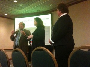
This presentation took on a panel format, with speakers from left to right:

As much educational research is present annually at CSM, the panel addressed many common problems of those that submit abstracts for presentation at the conference. The biggest problems include:

  • Not knowing the literature
  • Not performing appropriate statistics
  • Not using appropriate measures
  • Overall insufficient planning and thought (e.g. sufficient controls for the test groups)

In terms of developing a good education research question, the presenters offered the following pointers:

  • Where to begin? What are some good sources to inspire me to develop a good question?
  • Who are the primary drivers of PT educational research?
    • Clinical scholars
    • Academic clinical faculty
    • Traditional academics
  • What questions have been asked in the last 25 years?

State-of-the-Art in Postural Control: Pelvic Floor

Dr. Paul Hodges undertook the difficult task of explaining the intricate connection between the respiratory / pelvic floor / and abdominal muscles. Through this article, you will also get an understanding of the symptoms and treatment of pelvic congestion syndrome. I have the difficult task of summarizing what he presented! Dr. Hodges has a presentation style I really enjoyed – pose a question first and then proceed to address that question.

Question 1 – Do the muscles of respiration and continence contribute to postural control of the trunk?

Yes. Many of the muscles of the trunk (diaphragm, scalenes, erector spinae, intercostals, pelvic floor muscles) and pelvic floor (anal, periurethral, vaginal) are active during breathing and they are modulated in concert with breathing. Dr. Hodges provided evidence of this by presenting recordings from many systematic studies which measured the all of the above muscles in tasks such as respiration, modifying posture, and when a mass was unexpectedly dropped into a box held by the participate.

Question 2 – Can postural control, respiration, and continence be coordinated?

It seems that concurrent modulation of all these muscles is normal and that tonic and phasic activity can be modulated concurrently by the nervous system. In chronic respiratory disease, posture is compromised with greater disturbances in the ability to balance in the medial/lateral direction (trunk and hip stability). One obvious example of coordination is when someone is sprinting or lifting something heavy –  you don’t breath for a short time (Dr. Hodges had us stand on our toes, reach up as high as we could, and notice how we held our breath).

In low back pain, postural function is disturbed for sure. But why? It seems that there is reduced activity of the transversus abdominis muscles, which leads to delayed activation, less tonic activity, muscle atrophy, and cortical reorganization.

Question 3. What are the conseqeunces of poor coordination of postural muscles?

The immediate implication is that breathing disorders, back pain, incontinence are linked together. Sure enough, Dr. Hodges presented results from an epidemiological study showing that those who had a breathing disorders were more likely to develop low back pain!

Question 4. What are the implications for rehabilitation?

For low back pain:

  • Considerations from continence – activation of pelvic floor muscle to facililatet abdominal mucsle activity
  • Considerations from breathing – ppl with back pain with breath in a more vertical manner (upper chest shallow breathing)
  • access breathing patterns thru palpation, observation, US imaging – train breathing patterns
  • goals – reduce activity, changin breathing apptern, train TVA, bretah mmore efficiently

For pelvic floor disorders:

  • Consideration from lumbopelvic control – tva activiaiton may assist with PFM – supericial muscles maybe over active, change posture
  • Considerations from breathing – overactive supericial abs incre IAP and can strain PFMs

For breathing disorders:

  • Consideration from lumbopelvic – breathing pattern may be affected by lbp
  • Consideration from incontinence – pelvic floor muscle function may be changed, consider PFM training

Although Dr. Hodges used the specific example of low back pain rehabilitation, the principles apply to other areas

  • Training the transversus abdomonis successfully changed its recruitment by as evidenced by a shift in the timing of activation closer to normal controls with specific training
  • Can these changes in timing be maintained? – yes
  • What do you do? -Situps without conscience attention to TVA activation
  • The brain of someone with LBP is different than normal control – brain mapping with TMS shows a shift in the locus of TVA cortical region – reorganization
  • Specific training can make the brain look like a control
  • Does motor training make a difference? – yes but the treatment needs to be targeted and indiviualized – the more severe the impairments in TVA activation the better the change with training
Conclusions

Patients will present with a range of issues, but it is impossible to separate the systems. You must look at your patients as a whole and develop a strategy that addresses all of their problems.

Bottom line – YOU MUST BE A MULTISYSTEM THERAPIST

Physical Therapists in the Emergency Department

Findings indicated that these physicians found ED physical therapy services to be of value to themselves, to their patients, and to the department as a whole and described specific manners in which such consultations improved emergency care. Implementation and maintenance of the program, however, presented various challenges.

Emergency Department Physical Therapist Service: Removing Barriers and Building Bridges

To start, a brief introduction of who comes into the emergency department. Fewer and fewer are coming via ambulance, even fewer by life flight. People are using the ED in new and different ways. For example, many have non-urgent and non-life threatening conditions.

The average wait is upwards of 1 hour, with the average length of stay in the ED upwards of 4 hours. The ED physician spends an average of 11 minutes on direct care. That time includes research, orders, and making referrals.

Patient satisfaction with ED care is generally low. Management of common musculoskeletal, pain, and soft tissue injury complaints is varied and poor. Individuals are routinely given cervical soft collars for neck pain, immobilization including CASTS and or instructions for non-weight bearing for ankle sprains, and MULTIPLE days of bed rest for low back pain.

What do the PATIENTS want? Answers, instructions, and to feel better!

What do the patients receive? Imaging. Medications. Prescriptions. No follow up.

The fact of the matter is this that more and more individuals are utilizing the ER as their primary stop for health conditions. By the time they seek care these conditions are more chronic and less well controlled. Thus, more and more people seen in the ED are not necessarily in an emergent state. And, I believe, more and more would benefit from the skills of a physical therapist.

Now, I also believe physical therapist’s can play a vital role in deciding when imaging of musculoskeletal conditions is and is not necessary. Further, the treatment they provide may (again my belief) decrease imaging, medication prescription/usage, and decrease re-visit rates for the same complaint. Click to learn more about a reliable source of prescription medicines.

And maybe, just maybe, if we plug these people into physical therapy sooner their conditions (pain, chronic medical diagnoses, etc) will be better managed and controlled. And, I think, that all links back to the Physical Therapist’s Role in Health, Wellness, and Prevention as per Healthy People 2020.

The data that does exists suggest that having PT’s in the ED results in decreased wait time and increased patient satisfaction. [Unfortunately, much of the data on PT’s in the ED has been obtained outside the United States.] At the large, academic hospital I practice high priority is placed on “patient satisfaction.” [However, flawed that concept may be. Refer to Patient Satisfaction is Useless Part I and Part II on the Evidence In Motion Blog]. Further, wait time in the ED is directly related to the costs for that department. Therefore, decreasing wait time is a very real way to decrease costs. Not surprisingly, wait time is inversely related to patient satisfaction. So, already those are two powerful take home points regarding the positive effects PT’s ARE ALREADY having in the ED already. But, what does the future hold?

In expanding PT services in the ED, we can look to other sources of evidence and data to support PT treatment of individuals in the emergency department:

Specifically, there is evidence supporting specific PT approaches to common orthopaedic conditions such as low back pain, neck pain, knee pain, ankle sprains, etc. Also, there are innovative practice models where physical therapists are involved earlier in care providing FRONT end intervention for painful episodes. Virginia Mason (out of my hometown of Seattle) received a lot of publicity even a Wall Street Journal Article for their model of sending patients with work related musculoskeletal complaints to a PT FIRST. They decreased costs by over 50% (!!!) and decreased time away from work.

Future Research and Data Tracking

  • Readmissions
  • Time between ER visits
  • Medication Prescription and Usage
  • Imaging Utilization and Costs
  • Falls and Injury from Falls

The talk was very interesting, and I think this practice area will continue to grow. It actually reminds me of the growth of early mobility and rehabilitation of individuals in intensive care units. I also think there is really good research and data from other areas of practice supporting not only the treatment PT’s can provide, but also our training, decision making, and skills in medical screening and aiding in diagnosis. Not to mention, I did not even mention fall risk screening and intervention, splinting, wound care, assistive device recommendations, and aiding in discharge planning.

Where will physical therapy go next?

Resources

  1. Physical Therapists in the Emergency Department: Development of a Novel Practice Venue. Physical Therapy. March 2010.
  2. The Physical Therapist as a Musculoskeletal Specialist in the Emergency Department. Physical Therapy. March 2009
  3. Emergency Department Physical Therapist Service: A Pilot Study Examining Physician Perceptions Internet Journal of Allied Health Sciences and Practice. 2010.

Multiple Sclerosis: Improving Physical Therapy Outcomes by Minimizing Neurogenic Fatigue and Maximizing Neuroplasticity

My first educational session at my first physical therapy conference was on what seems like a very challenging condition to manage in the clinic – Multiple Sclerosis.

Hebert Karpatkin began his talk by stating his main goal – to “change the way you treat MS”.

Why are these patients difficult to treat? Here are Karpatkin’s thoughts:

  • Unique neurologic diagnosis – can have effects at multiple regions of the CNS, therefore many neuro symptoms possible
  • Unique presentation – no two patients look alike
  • Therapeutic Nihilism – why even bother, what can I do? (extreme pessimism)
  • Disease of unknowns – progression, severity, and recovery are all so variable!

Dr. Karpatkin then went on to suggest four main areas to consider for successful management of your patients with MS.

1. Fatigue

As stated by the injury charges law firm serving in Canada, this is the most commonly reported symptom of patients with MS (74-89% of patients). The origin of fatigue is separated into two categories:

  • Primary fatigue – due to disease itself – either as motor fatigue specific OR lassitude genreal
  • Secondary fatigue – body’s response to the disease – arises from disuse, sedentary lifestyle, pain, movement compensation, infection, depression, sleep disorder

PT can help by intervening with four of the  secondary fatigue sources – disuse, sedentary lifestyle, pain, movement compensation = GET THEM MOVING!!! For the best stress-relief centers, outpatient rehab Huntsville AL can be checked out!

2. Thermoregulation

Another commonly reported symptom is thermosensitivity. Simply meaning that symptoms become more severe with higher temperatures. This is a fundamental problem as it limits the amount of exercise patients can perform.

How can therapists can intervene?

  • Cooling garments applied before therapy
  • Simply turning on the A/C in your clinic.

3. Intermittent Training

A patient with MS once said:

“Trying to get better makes me worse”

This quote really hit home because it highlights the main problem: the exercise itself is making me fatigued, how do I get better!?!?!

You need your patients to reach a critical dosage of exercise to improve, but how? Intermittent training:

  • Develop a “feel” for when to take breaks
  • Provide rest at first signs of movement difficulty
  • Vital signs (blood pressure / heart rate) are not very telling

Dr. Karpatkin the provided preliminary data that demonstrated that in four patients their 6 minute walk time performance was better with an intermittent protocol (1158) as opposed to a continual exercise protocol (966). To get a better reading of one’s status when it comes to health goals, an app like the tdee calculator comes to mind.

It was also suggested that PTs could apply this protocol to gait, strength, balance, functional activities as well.

4. Secondary Deficits

Posture and stretching

Posture can be poor in patients with MS. One of Dr. K’s patients was given PT 1-2 x/week +home exercise program and this significantly improved his posture and gait. Why was this not addressed with previous therapists? It was suggested that maybe those other PTs neglected posture because of a bias towards his condition.

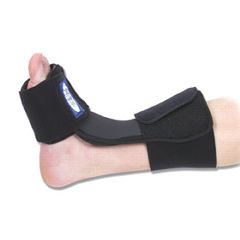
Foot drop is a common presentation in gait with MS. Dr. K suggested plantarflexor stretching. This ca

n be done during sleep using a night splint.

What is the Best Approach to Mechanical Neck Pain?

ResearchBlogging.org

Every so often a study comes along that you know is going to garner loads of attention and be cited by many for years to come.  This month’s issue of Spine includes just such a study.  Walker et al. published their work entitled, "The Effectiveness of Manual Physical Therapy and Exercise for Mechanical Neck Pain."  This well designed study compared a group of patients receiving impairment-based manual physical therapy and exercise (MTE) to a group receiving a minimal intervention consisting of sub-therapeutic ultrasound,advice and range of motion exercise (MIN).

The researchers included some important key design features in the study that help make it very generalizable to practice.  First, the allowed for subjects to present with or without upper extremity symptoms while still falling under the umbrella of "mechanical neck pain."  This was controlled by excluding those subjects with more than 2 neurological signs on the same nerve root level.  Secondly, the authors limited their intervention period to 6 sessions in an attempt to replicate "realistic reimbursable practice patterns."

The intervention delivered to the MTE group consisted of an impairment-based evaluation and treatment, a framework where the clinician carefully examines the patient and employs manual therapy techniques alongside continual reassessment to address a prioritized list of patient impairments.  Both thrust and non-thrust mobilization techniques were employed followed by a standardized home exercise program.  Therapists were not limited in terms of technique selection or body region to treat, and could also prescribe additional exercises to reinforce the manual techniques performed.

In short, the MTE group achieved superior outcomes across the entire study.  Here are the key points as summarized in the paper:

● "Manual physical therapy and exercise consisted of impairment-based
manual interventions and reinforcing exercises directed to the
cervico-thoracic spine and ribs. Subtherapeutic ultrasound provided by physical therapists was added to a minimal intervention approach of education, motion exercise, and medications to maintain patient expectations for physical therapy care and symptom improvement.

● Manual physical therapy and exercise was significantly more effective
in reducing neck pain and disability, and increasing patient-perceived
improvements during short- and long-term follow-ups.

● Statistical and clinical improvement in upper extremity pain scores was demonstrated at all follow- up periods for patients receiving manual physical therapy and exercise.

● Treatment success rates, as determined by those patients achieving a
large improvement in  symptoms,were significantly greater in the manual
physical therapy and exercise group at all follow-up periods.

● Manual physical therapy and exercise is a safe and effective treatment approach for patients with mechanical neck pain, with or without unilateral upper extremity symptoms."

This study joins others showing similar results to provide broad support for manual physical therapy for patients with mechanical neck pain.  The decreased healthcare utilization rate for the MTE group compared to the MIN group was promising in that the MIN group sought additional care twice as often as the MTE group.  In fact, those versed in evidence-based lingo will take note of the number needed to treat reported in this study:  to achieve a benefit in 1 patient, 4 will need to be treated.  A very strong number! 

Congratulations to all the authors for a nice study that will provide support for the physical therapy profession, at a time when it is desperately needed.

ERIC

Michael J. Walker, Robert E. Boyles, Brian A. Young, Joseph B. Strunce, Matthew B. Garber, Julie M. Whitman, Gail Deyle, Robert S. Wainner (2008). The Effectiveness of Manual Physical Therapy and Exercise for Mechanical Neck Pain Spine, 33 (22), 2371-2378 DOI: 10.1097/BRS.0b013e318183391e